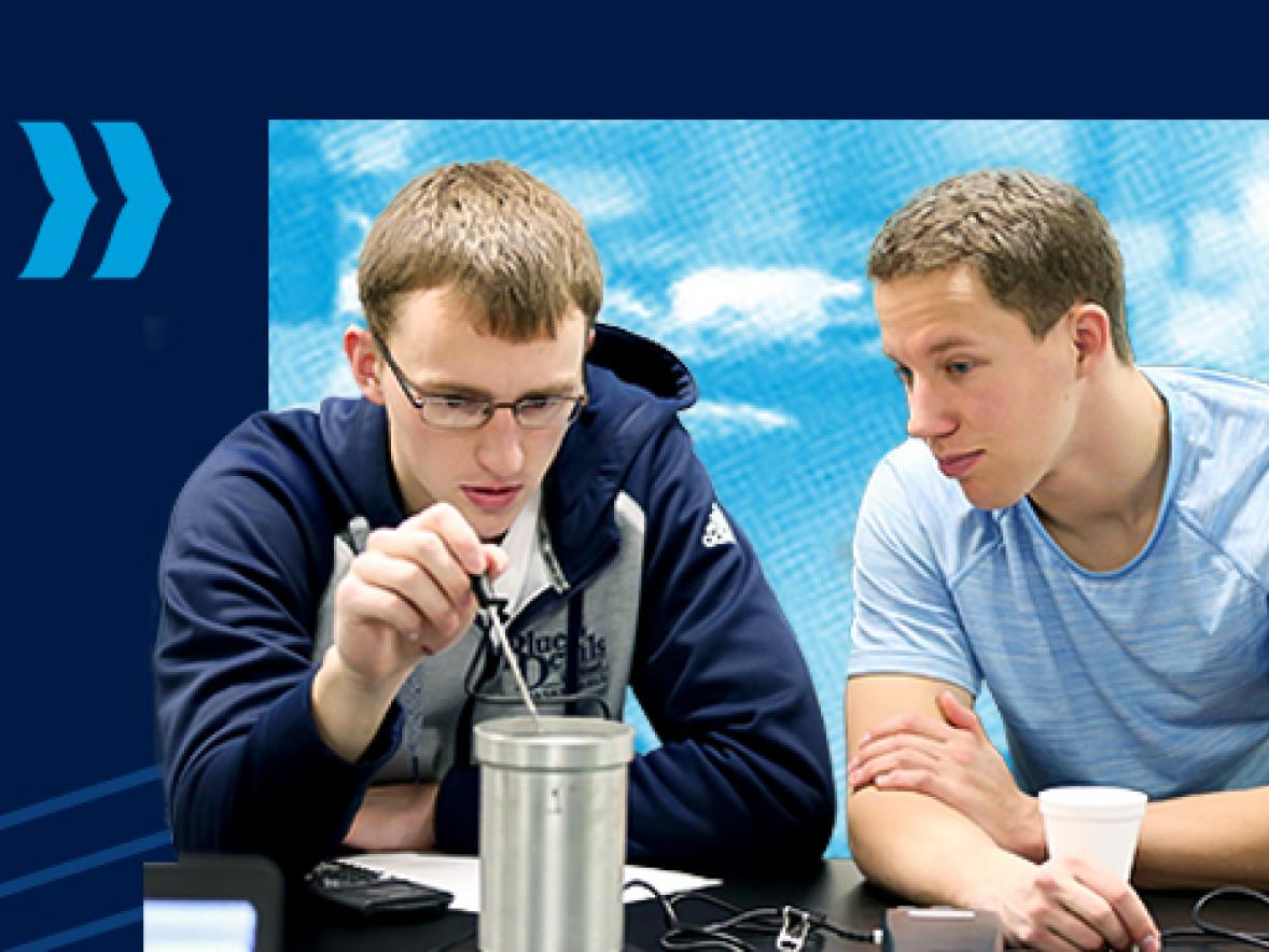
In a clean, temperature-controlled lab, six groups of students team up at stations in Lecturer Tiffany Hoage’s Anatomy and Physiology course in the Jarvis Hall Science Wing.
The lab is surprisingly splashed with color. Life-size human torso anatomy models contain removable pink plastic lungs, light-peach hearts, large gray intestines and other organs. Blue arteries and red veins run the length of the models like color-coded roadways.
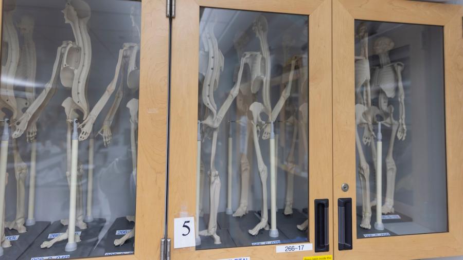
A colorful plastic skeleton, whose bones were painted in every color of the rainbow by Shelby Kaufer Saenger, a 2017 applied science alum, aids students’ memorization of the 206 bones in the human body and their features. A dozen miniature plastic skeletons line glass-fronted cabinets, like curious dolls on parade.
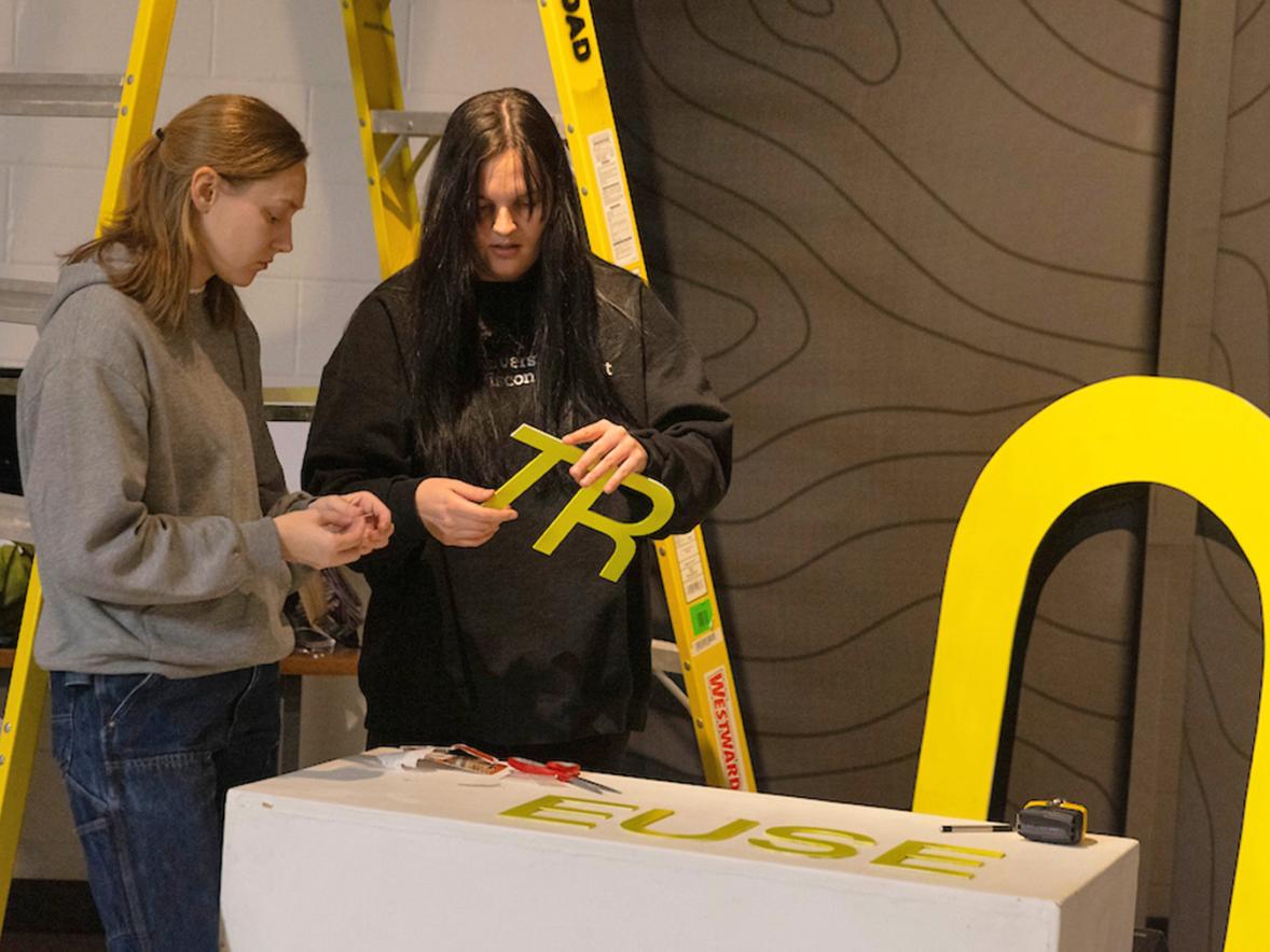
You immediately see the strange beauty, function and organization of the interior of the human body. The scratchy, zipper-like sound of Scotch tape ripping from rolls echoes across the room as students are busy labeling “body parts” that line the lab station counters.
Students in Anatomy and Physiology typically pursue Bachelor of Science degrees in applied biochemistry and molecular biology; applied science; dietetics; or health, wellness and fitness. But being a general education class, the students in this class section represent 11 different programs.
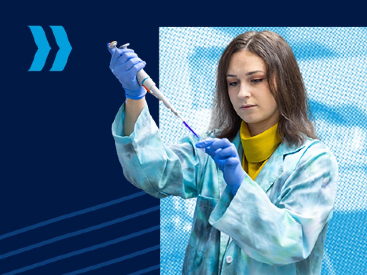
Students collect and stain their cheek cells and view them through microscopes at magnifications of up to 400X. For the osmosis and diffusion experiment, they place an artificial cell containing starch solution into an iodine solution to see how concentrations affect movement across the cell membrane.
“They’re exploring the world of cells. The cheek cells look like eggs in a frying pan. The ‘yolk’ is the nucleus and appears yellow because of the iodine stain,” Hoage explained.
“In the osmosis and diffusion experiment, water will go into the cell via osmosis, causing the cell to swell. Iodine will enter the cell via diffusion and react with the starch to produce a blue color. The concept of osmosis is important because if an IV solution does not contain the appropriate saline concentration, red blood cells can swell and burst or shrivel, respectfully,” she added.
A student jots notes in a notebook with a pattern of insects adorning the cover. Teams use the cell figure they labeled before the lab to identify the parts of the plastic cell models. As they stick laminated labels to the cell models, the students quiz each other: the mitochondria produce energy via cellular respiration; the hair-like cilia move things, like debris in the respiratory tract; ribosomes synthesize proteins; the nucleus is the cell’s control center; and more.
For the tattoo activity, students use anatomical terms and tape measurers to determine tattoo locations. And in the sectioning activity, they sculpt models of the human head and face using small chunks of blue, green, red and yellow modeling clay. They mold the clay in the palms of their hands and shape miniature cheek bones, eyes, mouths, ears and the bridge of the nose – each face unique to its designer.
“It’s not very good,” jokes Lacey Mader, holding up her creation before sectioning it through the anatomical planes.
Mader, of Buffalo, Minn., and Kael Lamb are juniors in health, wellness and fitness, and are interested in working with athletes in their careers.
“I took this class to learn more about the human body and how it functions. I’m excited that it will eventually help me in my internship and future job,” said Mader, who wants to work in athletic therapy.
Lamb, of Plainview, Minn., is on the Blue Devils football team. He’s hoping to intern at ETS, an alumni-owned training center, and train athletes as a career. “The hands-on experiences give me a deeper understanding of how the body works and moves, more than just terms and words to memorize,” he said.
